√ paranasal sinuses diagram 251981-How to treat paranasal sinuses
May , 13 · The paranasal sinuses are airfilled extensions of the nasal cavity There are four paired sinuses – named according to the bone in which they are located – maxillary, frontal, sphenoid and ethmoid Each sinus is lined by a ciliated pseudostratified epithelium, interspersed with mucussecreting goblet cellsNinja Nerds,Join us in this video where we discuss the anatomy of the sinus skull and go into detail on the different types of sinus cavities through the useStart studying Paranasal sinuses figure 911 Learn vocabulary, terms, and more with flashcards, games, and other study tools

Paranasal Sinuses Diagram Quizlet
How to treat paranasal sinuses
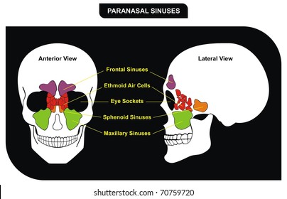
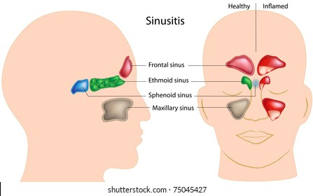
How to treat paranasal sinuses-Endoscopic paranasal sinus surgery The most important anatomic variations of the main paranasal sinus and accessory paranasal sinus Article in German KrmpotićNemaníc J(1), Vinter I, Jalsovec D Author information (1)Anatomisches Institut Drago Perovíc, Medizinische Fakultät Zagreb, KroatienOur paranasal sinuses come in pairs, with one of each pair located on either side of the face You have four of these pairs Sinus Anatomy – Paranasal Sinuses Maxillary Sinuses These are found directly in behind the check Ethmoid Sinuses These are smaller pockets of air, often described as having the appearance of honeycomb


Paranasal Sinuses Diagram Quizlet
Jan 13, 15 · These sinuses collectively are called the paranasal sinuses The name sinus comes from the Latin word sinus , which means a bay, a curve, or a hollow cavity Picture of the sinusesREAD MORE BELOW!In this video, we explore the 4 paranasal sinuses and what their proposed functions areINSTAGRAM @thecatalystuniversityI'm starting an InsWebMD's Sinuses Anatomy Page provides a detailed image and definition of the sinuses including their function and location Also learn about conditions, tests, and treatments affecting the sinuses
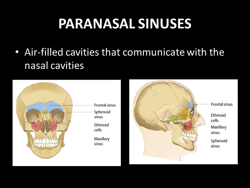
Paranasal Sinuses Diagram Quizlet Upgrade to remove ads Only $299/monthParanasal sinuses are air filled cavities in the frontal maxilae ethmoid and sphenoid bones From the nasal mucosa and their continued communication with the nasal fossae The largest sinus cavities are about an inch across 52 6 maxillary sinus see figThe paranasal sinuses are airfilled cavities in the frontal, ethmoid, maxilla, and sphenoid bones They're lined with a mucosal membrane and have small openings into the nasal cavity Maxillary sinus This sinus is located in the body of the maxilla behind the cheek just above the roots of the premolar and molar teeth
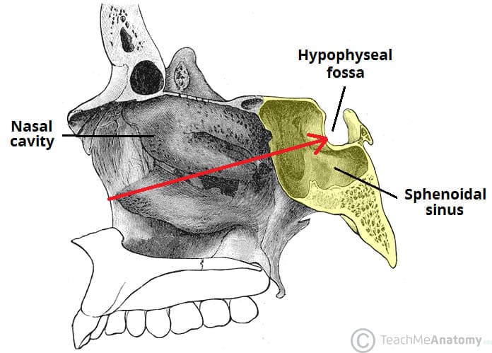
Start studying 2 Paranasal Set (Sinuses) Learn vocabulary, terms, and more with flashcards, games, and other study toolsParanasal Sinuses Paranasal sinuses refer to a group of airfilled spaces around the nasal cavity (a system of air channels that connect the nose with the back of the throat) (1)They facilitate the circulation of the air breathed in and out of the respiratory system (2) Paranasal sinuses have four different pairs maxillary sinuses, frontal sinuses, sphenoidal sinuses, and ethmoidalJun 05, 16 · Anatomically, the paranasal sinuses are in close proximity to the anterior cranial fossa, the cribriform plate, the internal carotid arteries, the cavernous sinuses, the orbits and their contents, and the optic nerves as they exit the orbits 31 – 35 The surgeon should be cautious when maneuvering instruments in the posterior direction to


21 2a Nose And Paranasal Sinuses Medicine Libretexts


Label The Diagram Below Using The Following Terms Chegg Com
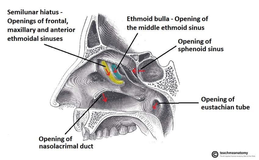
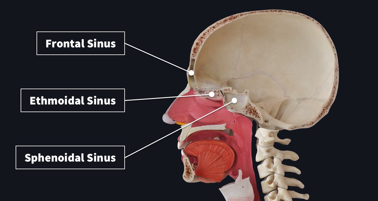
Paranasal sinuses organ included in Respiratory System PARANASAL SINUSES ANATOMY Air cavities, which in the nasal cavity are paired, between certain bones in the skull are known as paranasal sinusesThe bones which surround these sinuses and create the cavity are also responsible for the naming of the particular sinusDec 29, 12 · The sphenoethmoidal recess is located posteriorly,and not visible on this diagram Openings into the Nasal Cavity One of the functions of the nose is to drain a variety of structures Thus, there are many openings into the nasal cavity, by which drainage occurs The paranasal sinuses drain into the nasal cavity The frontal, maxillary andApr 19, 21 · The earliest stage of nasal cavity and paranasal sinus cancers is stage 0, also known as carcinoma in situ (CIS) The other stages range from I (1) through IV (4) Some stages are split further, using capital letters (A, B, etc) As a rule, the lower the
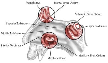


Functional Endoscopic Sinus Surgery Overview Preparation Technique


Paranasal Sinuses Illustration Anatomical Justice
RHINOLOGY (NOSE AND PARANASAL SINUSES DISORDERS) Nose and sinus issues are often associated with sinusitis, as well as other disorders, such as asthma, allergic and nonallergic nasal obstruction, nasal polyps, headaches and various types of allergies Alsero Travel offers highest level of care to patients with full range of sinonasal disordersLentworth LN, Krohel GB, et al Sinus tumor invading the orbit Ophthalmology 1984;919 Levin LA, Avery R, Shore JW, et al The spectrum of orbital aspergillosis a clinicopathological review Surv Ophthalmol 1996; Ogle OE, Weinstock RJ, Friedman E Surgical anatomy of the nasal cavity and paranasal sinusesAug 17, 16 · DEVELOPMENT OF PARANASAL SINUSES PNS appear as diverticula from nasal cavity Maxillary and sphenoidal sinus begins to develop before birth Frontal and ethmoidal sinuses develop after birth Enlargement of paranasal sinuses is associated with overall enlargement of the facial skeleton 48


The Paranasal Sinuses Trialexhibits Inc
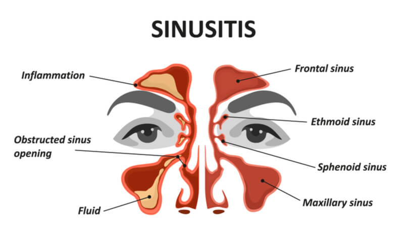


Maxillary Sinus The Definitive Guide Biology Dictionary
Mar 07, 16 · The paranasal sinuses are airfilled cavities within the ethmoid, frontal, and sphenoid bones and within the maxilla (Fig 175) They serve as resonating chambers for the voice and help to warm and moisten inhaled air The sinuses develop during childhood and are not fully formed until age 16 to 18Paranasal Sinuses STUDY Learn Flashcards Write Spell Test PLAY Match Gravity Created by erika_aranda PLUS Key Concepts Terms in this set (18) Innervation and blood supply to Frontal Sinus Supraorbital Nerve and Artery and Ethmoidal Arteries Frontal Sinus opening into theThe paranasal air sinuses often cause pain or discomfort if they get infected Let's have a look on anatomical models and a human skull and see if we can fin
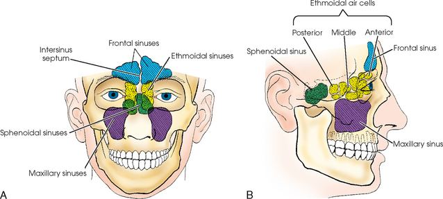


Paranasal Sinuses Radiology Key


Paranasal Sinuses Wikipedia
Apr 28, · Paranasal Sinuses Features Paranasal sinuses are airfilled spaces present within some bones around the nasal cavities The sinuses are Frontal, maxillary, sphenoidal, and ethmoidal All of them open into the nasal cavity through its lateral wall The function of the sinuses is to mack the skull lighter and add resonance to the voice In infections of the sinuses orParanasal sinus disease (sinusitis and mucoceles) may cause either an acute optic neuropathy plus pain on eye movements, similar to optic neuritis, or a chronically progressive optic neuropathy (Fig 537) The optic nerve disturbance results from compression or inflammation from a nearby mucocele501–503 In other cases contiguousThe paranasal sinuses are supplied by various branches of the internal and external carotid artery including the maxillary artery, anterior and posterior ethmoidal arteries, ophthalmic artery, supraorbital and supratrochlear arteries Share Improve this answer Follow


Amazon Com Emvency Wall Tapestry Sinuses Of Nose Human Anatomy Sinus Diagram The Nasal Cavity Bones Paranasal Sinusitis It Is Inflammation Maxillary Decor Wall Hanging Picnic Bedsheet Blanket 60x50 Inches Home Kitchen


Figure 1 From Chronic Inflammatory Disease Of Na Sal Cavity And Paranasal Sinuses Semantic Scholar
The sinuses are cavities or air filled pockets near the nasal passage It is the most important part of anatomy of nose and nasal sinus The nose is the part of the respiratory tract that sits front and center on your face In bones behind your nose are your sphenoid sinuses The bony lateral wall is convoluted by the turbinates called superiorJan 22, 18 · The maxillary sinus is one of the four paranasal sinuses, which are sinuses located near the nose The maxillary sinus is the largest of the paranasal sinuses The two maxillary sinusesApr 29, 16 Explore Natalie StickneyShade's board "paranasal sinuses", followed by 140 people on See more ideas about paranasal sinuses, sinusitis, radiology


Paranasal Sinuses Diagram Quizlet
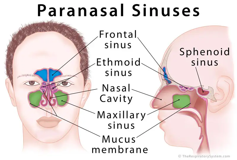


Paranasal Sinus Definition Location Anatomy Function Picture
Nose Diagram Image result for basic parts of the nose and their functions Saved by Lorna Omar 14 Nose Diagram Ear Congestion Sinus Drainage Paranasal Sinuses Nasal Septum Nasal Cavity Sinus Pressure The Voice Natural Remedies More information More like thisMay 28, · (a) Diagram of a coronal section through the frontal, ethmoid, and maxillary sinuses showing the osseous boundaries of the sinuses and their relationship to the nasal cavity and orbits (b) Diagram of a sagittal section through the nasal cavity, showing drainage pathways of the paranasal sinuses as they relate to the hiatus semilunaris andWhere do I get my information from http//armandohorg/resourceFacebookhttps//wwwfacebookcom/ArmandoHasudunganSupport me http//wwwpatreoncom/armando


The World S First Precise Model Of The Human Paranasal Sinus For Ess Training 03 03 27


Misunderstanding Sinuses Relaxed Face A Personal Journey Of Un Frowning
Paranasal sinuses Dr Sachintha Hapugoda and Dr Maxime StAmant et al The paranasal sinuses usually consist of four paired airfilled spaces They have several functions of which reducing the weight of the head is the most important Other functions are air humidification and aiding in voice resonance They are named for the facial bones inStart studying Figure 715 Paranasal sinuses Learn vocabulary, terms, and more with flashcards, games, and other study toolsThere are four groups of paranasal sinuses the maxillary, ethmoid, frontal, and sphenoid sinuses (see figure Locating the Sinuses) Sinuses reduce the weight of the facial bones and skull while maintaining bone strength and shape The airfilled spaces of the nose and sinuses also add resonance to the voice


Maxillary Sinus Cancer Tnm Stages And Grades Cancer Research Uk
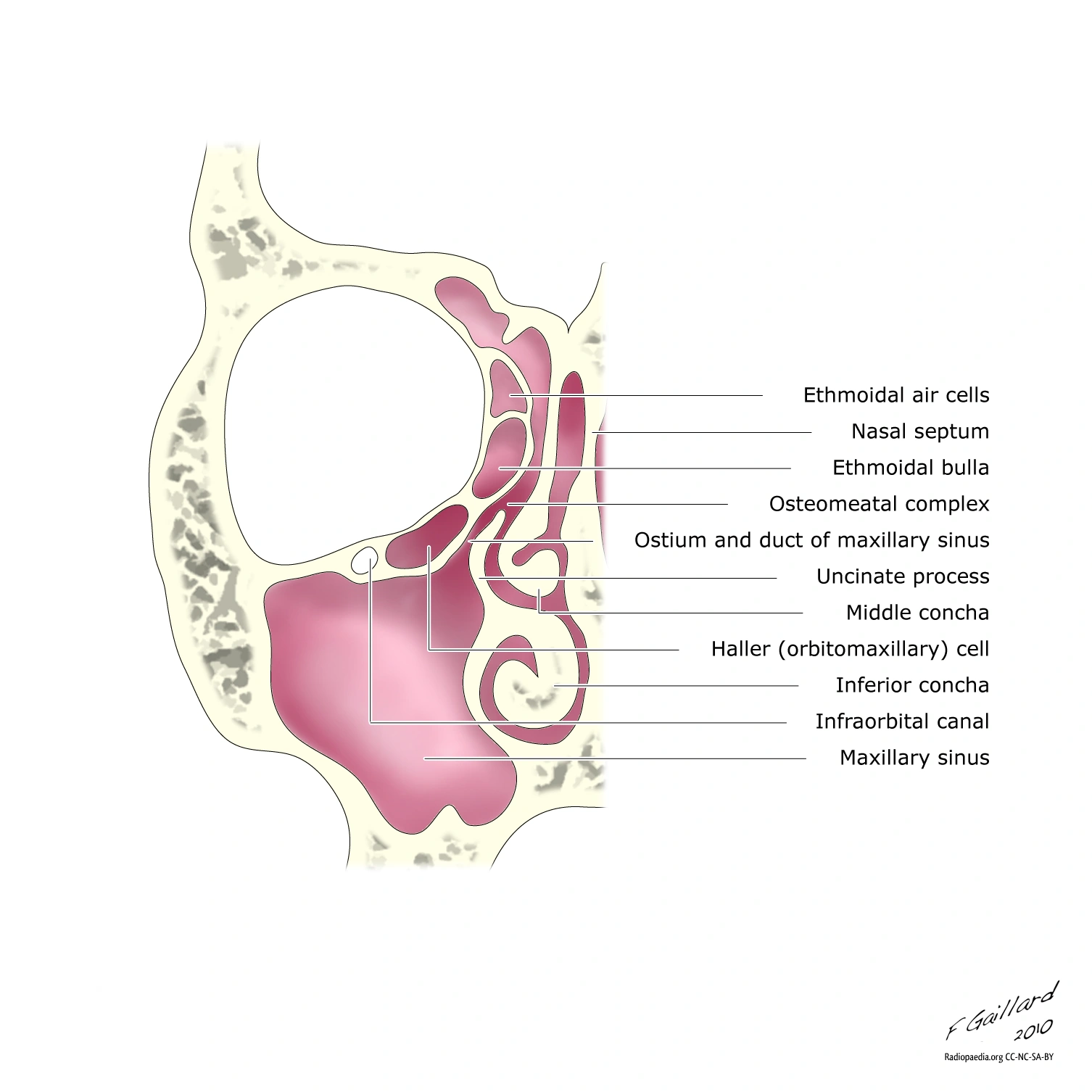


Nasal Cavity Paranasal Sinuses Bones Foramina Canals Ethmodial Cell Variants Ranzcrpart1 Wiki Fandom
Which start developing from the primitive choana at 25–28 weeks of gestation 1 Three projections arise from the lateral wall of the nose and serve as the beginning of the development of the paranasal sinuses The anterior projection forms the Agger nasi, the inferior orParanasal sinus anatomy The maxillary sinuses are the largest of the all the paranasal sinuses The nasal septum separates the left and right maxillary sinuses 9 In the frontal ethmoid and sphenoid of the cranium and the maxillary bones of the face Histology of the nose and paranasal sinusesMar 01, 17 · 1 Introduction The paranasal sinuses are group of air filled spaces surrounding the nasal cavity;
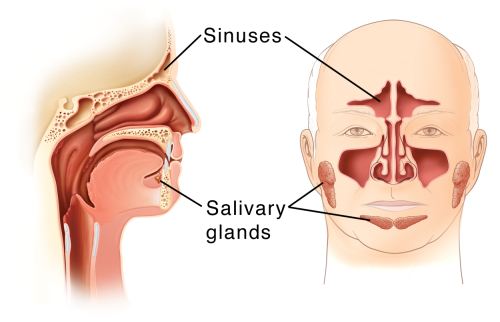


Paranasal Sinus Tumors University Hospitals


Sinus Diagram Stock Illustrations 337 Sinus Diagram Stock Illustrations Vectors Clipart Dreamstime
About Press Copyright Contact us Creators Advertise Developers Terms Privacy Policy & Safety How works Test new features Press Copyright Contact us CreatorsMar 16, 18 · Maxillary Sinus (within the maxillary bones) The largest among all the paranasal sinuses 2, these two conical cavities are located on the two sides of the nose, above the upper teeth, and below the cheeks 4 Ethmoid Sinus (within the ethmoid bones) Three to eighteen 5 air cells present in the ethmoid labyrinth, on both sides of the nose, between the eyes 6, 7May 31, 21 · The maxillary sinuses are the largest of the all the paranasal sinuses They have thin walls which are often penetrated by the long roots of the posterior maxillary teethThe superior border of this sinus is the bony orbit, the inferior is the maxillary alveolar bone and corresponding tooth roots, the medial border is made up of the nasal cavity and the lateral and anterior border


Startradiology


Nose And Paranasal Sinuses Structure And Function Organization Of The Respiratory System
Jan 27, 15 · Frontal sinuses The right and left frontal sinuses are located near the center of the forehead (frontal bone) just above each eye Maxillary sinuses These are the largest of the sinusesThe paranasal sinuses are joined to the nasal cavity via small orifices called ostiaThese become blocked easily by allergic inflammation, or by swelling in the nasal lining that occurs with a coldIf this happens, normal drainage of mucus within the sinuses is disrupted, and sinusitis may occur Because the maxillary posterior teeth are close to the maxillary sinus, this can also causeRoyaltyfree stock vector ID Sinuses of Nose Human Anatomy Sinus Diagram Anatomy of the Nose Nasal cavity bones Anatomy of paranasal sinuses Sinusitis It is the inflammation of the maxillary sinuses l


File Paranasal Sinuses Svg Wikimedia Commons


Nose And Sinus Anatomy Dr Thomas S Higgins Md Msph
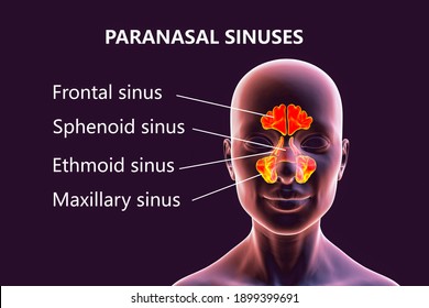
The paranasal sinuses include a maxillary sinus in each cheek six to 12 ethmoid sinuses on each side of the nose between the eyes a sphenoid sinus close to the center of the head behind the ethmoid sinuses and a frontal sinus on each side in the forehead Most individuals have four paired cavities located in the cranial bone or skull
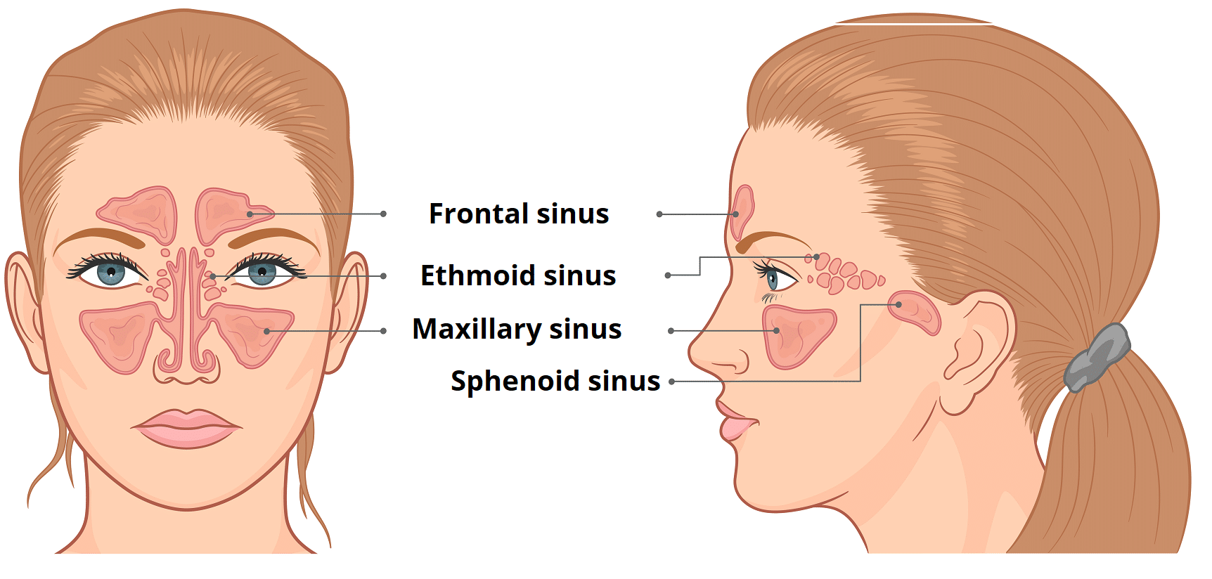


The Paranasal Sinuses Structure Function Teachmeanatomy


Nasal Cavity And Paranasal Sinus Cancer Miami Cancer Institute Baptist Health South Florida


Paranasal Sinuses And Sinusitis
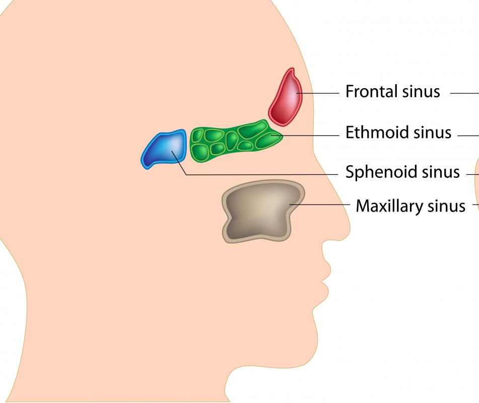


What Is A Sinus With Pictures
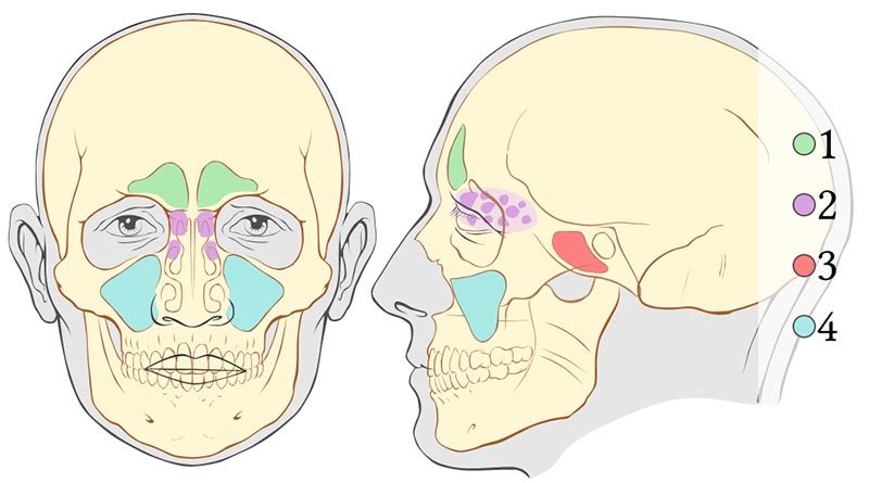


Sinus Barotrauma Divers Alert Network


Paranasal Sinuses Diagram Quizlet
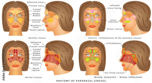


Nasal Sinus Nasal Sinus Human Anatomy Sinus Diagram Anatomy Of The Nose Nasal Cavity Bones Anatomy


Maxillary Sinus Radiology Reference Article Radiopaedia Org
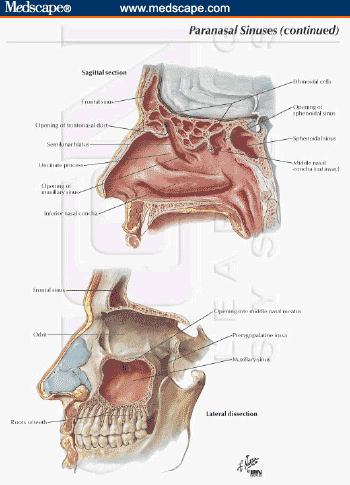


A Practical Approach To The Patient With Sinusitis


Paranasal Air Sinuses Location Functions Relations And Applied Anatomy Qa


Paranasal Sinuses
:background_color(FFFFFF):format(jpeg)/images/article/en/the-paranasal-sinuses/972PC0nYOzlz7wqSgLmNA_sinus_frontalis_large_u9Vfozc0uUoMtc6KtIaUfw.png)


Paranasal Sinuses Anatomy And Clinical Aspects Kenhub


Volumetric Surface Renderings Of Paranasal Sinus Created From Download Scientific Diagram


Paranasal Sinuses Maxillary Ethmoid Sphenoid Frontal Notes Youtube


Http Ksumsc Com Download Center Archive 1st 433 03 Respiratory block 433 teams work Anatomy L1 nose nasal cavity Pdf
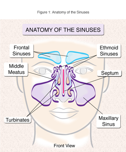


Sinusitis Treatment And Surgery Nyc


1


Startradiology


Pin On Nursing School


Paranasal Sinuses Images Stock Photos Vectors Shutterstock


Figure Paranasal Sinuses Frontal Sinus Ethmoid Statpearls Ncbi Bookshelf


Figure Anatomy Of The Paranasal Sinuses Spaces Between The Bones Around The Nose Pdq Cancer Information Summaries Ncbi Bookshelf


Parts Of The Throat Mouth Nose And Ears With Diagrams Macmillan Cancer Support


7 Paranasal Sinus Illustrations Clip Art Istock


What Are Your Sinuses And How Do They Work John E Mcclay Md


Paranasal Sinus


The Paranasal Sinuses Stock Photos And Images Agefotostock


Pin On Health Wellness


Overview Of The Drainage Pathways Of The Paranasal Sinuses A Download Scientific Diagram


The Paranasal Sinuses Structure Function Teachmeanatomy


1


The Paranasal Sinuses Structure Function Teachmeanatomy


Sinusitis Animation Youtube


Hb 7 Parts Of The Paranasal Sinuses Diagram Quizlet


3 677 Sinus Stock Vector Illustration And Royalty Free Sinus Clipart


Radiology Of The Nasal Cavity And Paranasal Sinuses Ento Key


Paranasal Sinuses Human Anatomy Organs


What To Do About Sinusitis Harvard Health


Sphenoid Sinus Images Stock Photos Vectors Shutterstock
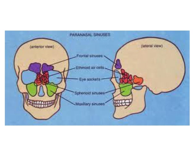


Paranasal Sinuses


Paranasal Sinus Anatomy What The Surgeon Needs To Know Intechopen
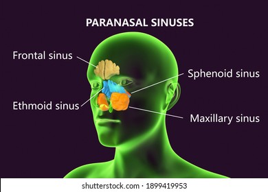


Sphenoid Sinus Images Stock Photos Vectors Shutterstock


1


Solved Directions Using Your Cpt Manual Key In The Corr Chegg Com


Sinuses Radiology Key
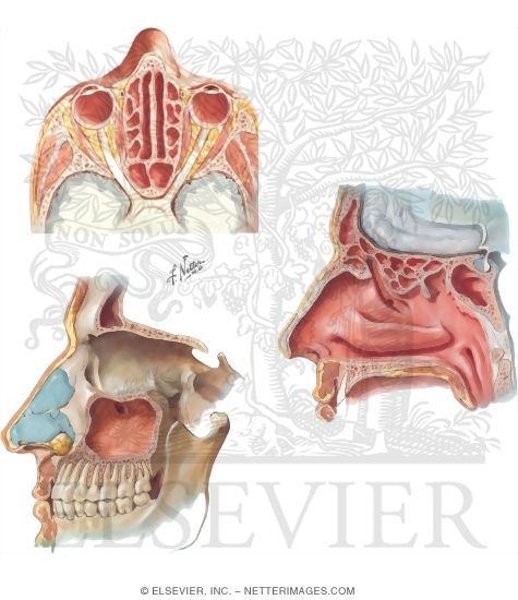


Paranasal Air Sinuses
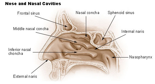


Seer Training Nose Nasal Cavities Paranasal Sinuses


Q Tbn And9gcsqidmgypqtou9zkiili2afvsssshl19qwomar6b1fzrmhxbxb7 Usqp Cau


Parts Of The Throat Mouth Nose And Ears With Diagrams Macmillan Cancer Support
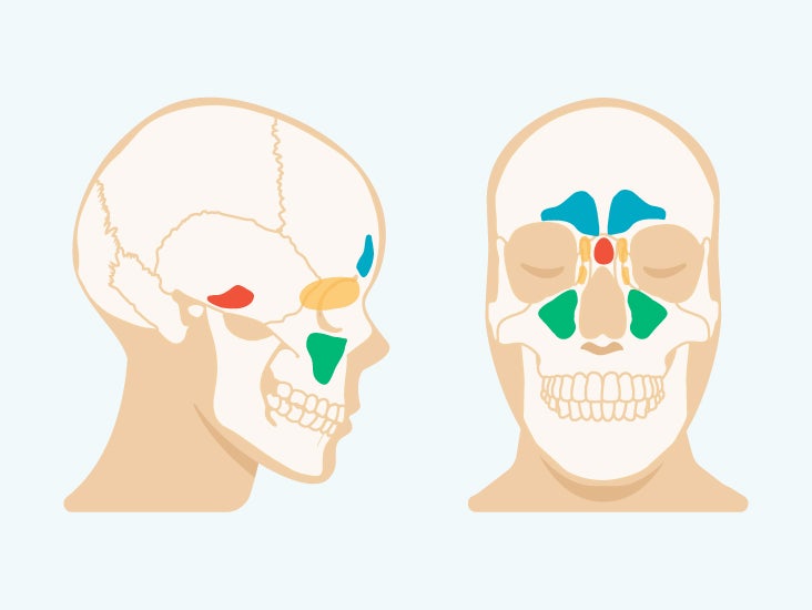


Sinus Cavities In The Head Anatomy Diagram Pictures


Paranasal Sinuses Wikipedia


Diagram Of Paranasal Sinuses Download Scientific Diagram
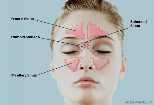


What Are The Sinuses Pictures Of Nasal Cavities


Paranasal Sinuses Diagram Quizlet


The Paranasal Sinuses Structure Function Sciencekeys


Anatomy Of The Sinuses Otolaryngology Houston


What Is Nasal Cavity Nose Cancer What Is Sinus Cancer


Location Of Paranasal Sinuses Download Scientific Diagram
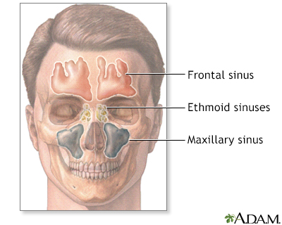


Sinus X Ray Information Mount Sinai New York


Paranasal Sinuses And Sinusitis


Paranasal Sinuses Diagram Quizlet


Pin On Glandular Odontogenic Cysts


Www Thieme Connect De Products Ebooks Pdf 10 1055 B 0036 Pdf


Pin On Health Tips


7 Paranasal Sinus Illustrations Clip Art Istock
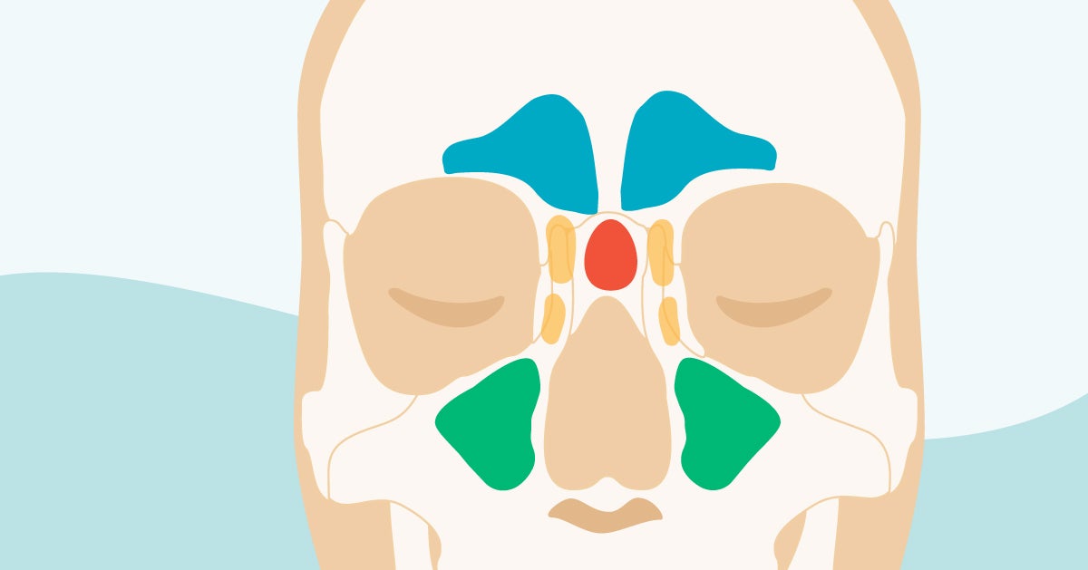


Sinus Cavities In The Head Anatomy Diagram Pictures


Paranasal Sinuses And Facial Bones Radiography Radiology Reference Article Radiopaedia Org


Disorders Of The Paranasal Sinuses In Horses Horse Owners Merck Veterinary Manual


Anatomy Of The Pharynx Diagram


Anatomy Of Nose And Paranasal Sinus By The


Paranasal Sinuses Images Stock Photos Vectors Shutterstock


Paranasal Sinus High Res Illustrations Getty Images


Facial Sinuses Anatomy Anatomy Drawing Diagram


Paranasal Sinus And Nasal Cavity Cancer Wikipedia


Paranasal Sinuses Anatomy Physiology And Diseases Ppt Video Online Download


Nose And Sinuses Ear Nose And Throat Disorders Merck Manuals Consumer Version


Paranasal Sinus Anatomy What The Surgeon Needs To Know Intechopen


Which Of The Bones That Contain Paranasal Sinuses Is Are Facial Bone S Rather Than Cranial Bone Lifeder English


About The Sinuses Michael R Anderson Md


Paranasal Sinuses Complete Anatomy


Nasal Sinus Anatomy Anatomy Drawing Diagram


The Nasal Cavity And Paranasal Sinuses Canadian Cancer Society


コメント
コメントを投稿